Dr. Tushar Deore is dedicated to providing the best care for Hip replacement.
 Total Hip Replacement
The hip is one of the largest weight-bearing joints. It consists of two main parts: a ball (femoral head) at the
top of the thighbone (femur) that fits into a rounded socket (acetabulum) in the pelvis. Bands of tissue called
ligaments (hip capsule) connect the ball to the socket and provide stability to the joint.
The bone surfaces
of the ball and socket have a smooth durable cover of articular cartilage that cushions the ends of the bones and
enables them to move easily. A thin, smooth tissue called the synovial membrane covers all remaining surfaces of
the hip joint.
In a healthy hip,
this membrane makes a small amount of fluid that lubricates and almost eliminates friction in the hip joint.
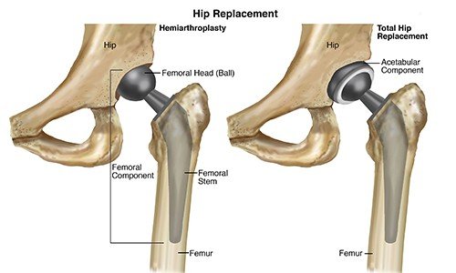 1. Acetabulum: The acetabulum is the cup-shaped part of the pelvis that forms the socket for the ball of the femur. In total hip replacement, the acetabulum is replaced with a metal or plastic prosthetic component.
2. Femoral Head: The femoral head is the ball-like structure at the top of the femur that fits into the acetabulum. In total hip replacement, the femoral head is replaced with a metal or ceramic prosthesis.
3. Articular Cartilage: The hip joint is cushioned by a smooth layer of articular cartilage. In total hip replacement, the damaged cartilage is removed, and the joint is replaced with a prosthetic implant.
4. Synovial Fluid: The synovial fluid in the hip joint lubricates the area, reducing friction. After total hip replacement, the prosthetic materials used also provide a smooth surface to maintain this function.
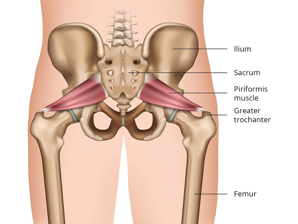
5. Muscles: The muscles surrounding the hip, such as the gluteus medius and iliopsoas, help stabilize and move the hip joint. After total hip replacement, rehabilitation focuses on strengthening these muscles to restore mobility and function.